Bulbar Strictures
Home > Urethral Stricture > Bulbar Strictures
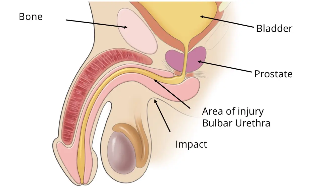
It is particularly susceptible to straddle trauma during sports, from straddling the bar of a bicycle, or from other straddle trauma such a skateboard misadventure or being kicked or hit with a ball.


The impact of the straddle trauma can cause the bulbar urethra (bulbous urethra) to be crushed directly against the surrounding bone. In some cases, the trauma can cause immediate swelling and the appearance of blood at the urethral opening at the tip of the penis. However, in many cases, a bulbar urethral stricture develops slowly over time, caused by the development of a scar and the growth of scar tissue as the injury heals. The oftentimes slow development of a stricture in the bulbar urethra means that some patients may not even remember the specific injury that led to the stricture. A bulbar stricture caused by straddle trauma is typically short in length, making treatment highly curable if properly performed.
The images below are examples of a bulbar urethral stricture as seen on a RUG, a type of diagnostic imaging for urethral strictures.
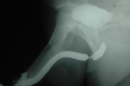
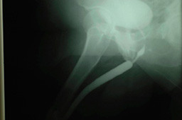
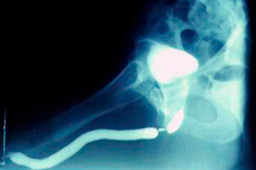
Before and After Imaging
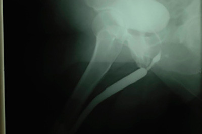
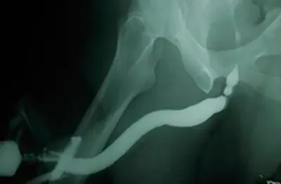
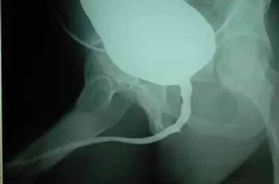
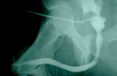
When a bulbar stricture of the urethra exceeds 1.5-2 cm in length or is recurrent, internal urethrotomy is an option with a very low success rate it “success” is defined as a permanent solution and not temporary improvement. The best initial treatment option for longer bulbar urethral strictures is open urethral reconstruction, also called urethroplasty. When the stricture is longer, as shown in the example below, excision and primary anastomosis is not a good treatment technique because too much of the urethra would need to be removed. This surgery is also called anastomotic urethroplasty. The resulting gap created if a longer bulbar stricture was removed would be too great for the ends to be re-connected without tension. Below are a few examples of bulbar urethral strictures longer than 4 cm in length that can not be treated using excisional repair.
Long Bulbar Strictures
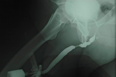
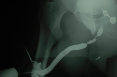
When the bulbar stricture of the urethra is too long for excisional repair, our preferred approach is to cut open the urethra and add buccal mucosa (a graft taken from the inside of the cheek) to augment and widen the narrowed area of the urethra. Regardless of the technique used or the location of a bulbar stricture, it can usually be repaired with a very high success rate and a low risk of complication.
We recently published our long-term results for the repair of bulbar urethral strictures with anastomotic urethroplasty and buccal mucosa graft onlay repair at the Center for Reconstructive Urology dating back to 1998.
Our protocol has always been to gently look inside the urethra with a scope 4 months after surgery with the use of a monitor to see if the urethra is wide open or not to assess early technical success. Our results with anastomotic urethroplasty was 100% and with (dorsal) buccal graft repair it was very 97.5%. Long-term success was 99.3% with anastomotic urethroplasty and 95% and buccal graft reconstruction.
Although bulbar stricture surgeries are considered by some Urologists to be “simple” or “bread and butter” repairs, there are published papers by experts who report an 85% or lower success rate. When this surgery is successful, the patient is cured. When the surgery fails, the problem is more complicated. However, over the past 20+ years, we have also performed many re-do repairs with a very high success rate.

